MORE...
We can SEE more for the simple reason that we invested in technology so we can discover conditions in early stages of development.
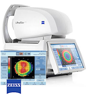
EYE PROFILE
The profile takes the eye’s “finger print” so we can understand problems like blurry vision, night vision and diseases of the cornea.
Typically used during EXAM
EYE MAP
We use Atlas topography to map the surface of the eye, detect potential diseases, and to design precise custom lenses.
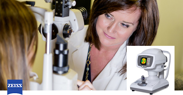
Typically used during EXAM
High Definition Retina Scan
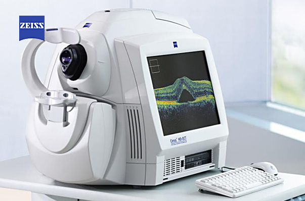
The Cirrus HD-OCT creates high resolution images of the eye. It allows us to see MORE of the eye in greater detail. We can detect diseases in their early stages.
Typically used during IN DEPTH DIAGNOSIS
Visual Field Mapping
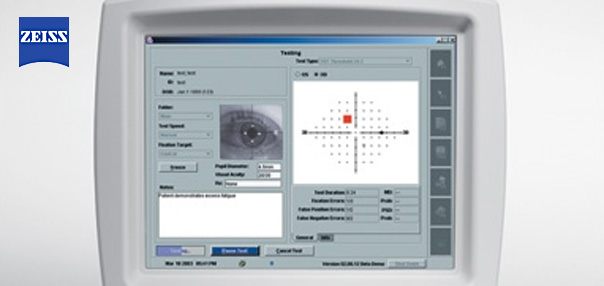
If you are a glaucoma patient we have you covered. The ZEISS Matrix technology is excellent for defining the status of the visual field and providing basic glaucoma monitoring.
Eye Imaging
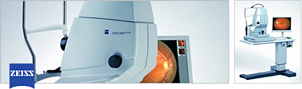
Retinal photography technology allows us to keep a detailed record of your eye condition and allows us to monitor for changes over time.